Which stain is used for Corynebacterium Diphtheriae?
Albert’s method, of staining diphtheria cultures consists of staining a fixed smear for one minute (some laboratories stain for five minutes) with a solution containing toluidine blue and malachite (or methyl) green, washing with water, and applying Albert’s iodine for one minute.
What is KLB in microbiology?
Corynebacterium diphtheriae is the pathogenic bacterium that causes diphtheria. It is also known as the Klebs–Löffler bacillus, because it was discovered in 1884 by German bacteriologists Edwin Klebs (1834–1912) and Friedrich Löffler (1852–1915).
What is the test for diphtheria?
Doctors usually decide if a person has diphtheria by looking for common signs and symptoms. They can swab the back of the throat or nose and test it for the bacteria that cause diphtheria. A doctor can also take a sample from an open sore or ulcer and try and grow the bacteria.
Which color is taken by Corynebacterium Diphtheriae after Gram staining?
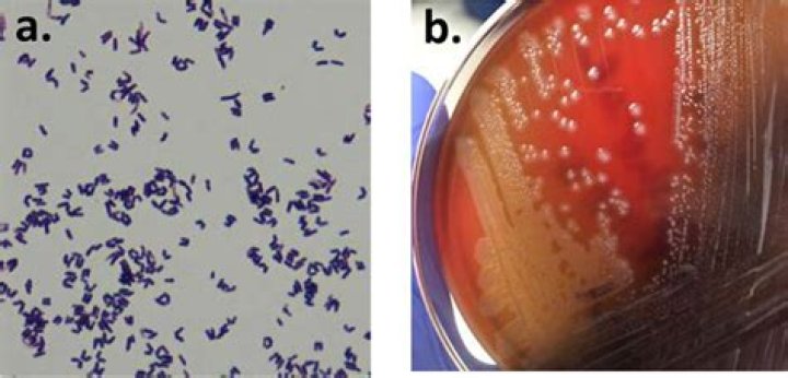
Corynebacterium Infections In a typical Gram stain these bacteria appear as V-in Y-shaped arrangements or in clumps that resemble Chinese letters. Corynebacteria are nonmotile, catalase positive, aerobic, and contain metachromatic granules when stained with methylene blue.
How do you stain diphtheria?
Introduction. Albert’s staining technique is a type of special staining technique since it is used to demonstrate a special structure in bacteria. It is chiefly used to demonstrate metachromatic granules found in Corynebacterium diphtheriae. This bacterium is responsible for the disease diphtheria.
What is Albert stain?
Albert stain is a type of differential stain used for staining high-molecular-weight polymers of polyphosphate known as metachromatic granules or volutin granules found in Corynebacterium diphtheriae. Metachromatic granules are also found in Yersinia pestis, and Mycobacterium species.
What is Pseudomembrane in diphtheria?
Within two to three days, the dead tissue forms a thick, gray coating that can build up in the throat or nose. Medical experts call this thick, gray coating a “pseudomembrane.” It can cover tissues in the nose, tonsils, voice box, and throat, making it very hard to breathe and swallow.
How do you stain Volutin granules?
Procedure
- Prepare a smear on clean grease free slide.
- Air dry and heat fix the smear.
- Treat the smear with Albert’s stain and allow it to react for about 3 mins .
- Drain of the excess stain do not water wash the slide.
- Flood the smear with Albert’s iodine for 2 minutes.
What is the differential diagnosis of diphtheria?
The differential diagnosis of diphtheria includes streptococcal pharyngitis, viral pharyngitis, Vincent’s angina, infectious mononucleosis, oral syphilis and candidiasis. If the infection involves the larynx, the patient can present with fever, hoarseness and a barking cough.
What are the five types of diphtheria?
Respiratory and cutaneous diphtheria are caused by toxic strains of the bacteria Corynebacterium diphtheriae and Corynebacterium ulcerans and very rarely Corynebacterium pseudotuberculosis….Diphtheria
- classical respiratory diphtheria.
- laryngeal diphtheria.
- nasal diphtheria and.
- cutaneous diphtheria (skin lesions).
What is Endospore staining in microbiology?
Endospores staining is the type of staining to recognize the presence spore in bacterial vegetative cells. The bacterial endospores need a staining which can penetrate wall thickness of spore bacteria. A method of endospores staining is Schaeffer Fulton method that used Malachite Green.
