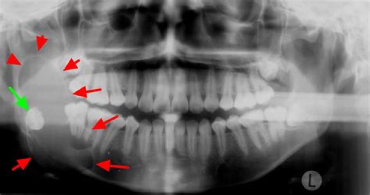
What is the classic radiographic appearance of the ameloblastoma?
According to Worth,[4] the most common radiographic appearance of ameloblastoma is a multilocular radiolucency with a corticated border, and margins, which usually show irregular scalloping.
How do you describe ameloblastoma?
Ameloblastoma is a rare, noncancerous (benign) tumor that develops most often in the jaw near the molars. Ameloblastoma begins in the cells that form the protective enamel lining on your teeth. The most common type of ameloblastoma is aggressive, forming a large tumor and growing into the jawbone.
What does an ameloblastoma look like?
Ameloblastoma is characterized by an abnormal growth in the sinus area or jaw, often at the site of the third molar. The tumors or cysts may be aggressive and may spread to the nose, eye socket and skull.
What is the content of ameloblastoma?
Ameloblastoma is a benign odontogenic tumor generally present in the jaw bone. The tumor originates from the residual epithelium of the tooth germ, epithelium of odontogenic cysts stratified squamous epithelium and epithelium of the enamel organ. It represents approximately 1% of oral tumors.
How can you tell the difference between ameloblastoma and Dentigerous cyst?
Although the presence of a tooth within a lucent mass is pathognomonic for a dentigerous cyst, the aggressive features of portions of the mass and the presence of solid enhancing nodular foci were inconsistent with this type of cyst. Thus, ameloblastoma was the primary differential diagnosis.
Why is it called ameloblastoma?
If an aggressive tumor is left untreated, it can obstruct the nasal and oral airways making it impossible to breathe without oropharyngeal intervention. The term “ameloblastoma” is from the early English word amel, meaning enamel and the Greek word blastos, meaning germ.
Is teeth vital in ameloblastoma?
Involved teeth are vital and not displaced by the presence of this cyst.
How can you distinguish between ameloblastoma and odontogenic Keratocyst?
Most OKCs showed smooth border and unilocular shape, while most ameloblastomas showed scalloped border and multilocular shape. Compared with ameloblastomas, OKCs showed greater frequency to be associated with impacted tooth, and were unlikely to cause tooth displacement and root resorption.
How is ameloblastoma treated?
Ameloblastoma treatment usually includes surgery to remove the tumor. Ameloblastoma often grows into the nearby jawbone, so surgeons may need to remove the affected part of the jawbone. An aggressive approach to surgery reduces the risk that ameloblastoma will come back. Surgery to repair the jaw.
