What is Perthes disease Radiology?
Perthes disease, also known as Legg-Calvé-Perthes disease, refers to idiopathic osteonecrosis of the femoral epiphysis seen in children. It should not be confused with Perthes lesion of the shoulder.
What is the lateral pillar?
The lateral pillar of the femoral head is an important site for disease development such as osteonecrosis of the femoral head. The femoral head consists of medial, central, and lateral pillars.
How do I know if I have Perthes disease?
The symptoms of Perthes’ disease include:
- an occasional limp in the earlier stages.
- stiffness and reduced range of movement in the hip joint.
- pain in the knee, thigh or groin when putting weight on the affected leg or moving the hip joint.
- thinner thigh muscles on the affected leg.
How is Perthes disease diagnosed?
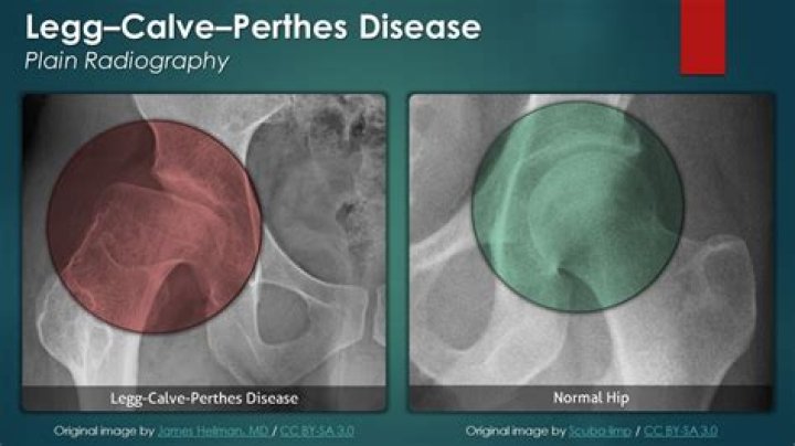
How is Perthes disease diagnosed? If your child is experiencing symptoms of Perthes disease, your healthcare provider will order an X-ray. X-rays are a common way to confirm a diagnosis. Your provider may also order additional studies, such as an MRI, to see how far the disease may have progressed.
Is Perthes painless?
Signs and symptoms of Perthes disease can include: walking with a limp (can be a “painless limp”) limited range of motion and stiffness in the hip, groin, thigh, or knee.
What type of disease is Legg-Calve-Perthes?
Legg-Calve-Perthes (LEG-kahl-VAY-PER-tuz) disease is a childhood condition that occurs when blood supply to the ball part (femoral head) of the hip joint is temporarily interrupted and the bone begins to die. This weakened bone gradually breaks apart and can lose its round shape.
How do you assess Legg-Calve-Perthes disease?
X-rays. Initial X-rays might look normal because it can take one to two months after symptoms begin for the changes associated with Legg-Calve-Perthes disease to become evident on X-rays. Your doctor will likely recommend several X-rays over time, to track the progression of the disease. MRI .
