What Is hip arthrography?
Arthrography is a type of imaging test used to look at a joint, such as the shoulder, knee, or hip. It may be done if standard X-rays do not show the needed details of the joint structure and function.
How would you perform hip joint arthrography?
Arthrography involves placement of a needle intra-articularly with aspiration of joint fluid and injection of a contrast agent to enhance imaging. It is often performed to exclude an infection in patients with hip pain or in patients with total joint prostheses.
What does a hip MRI arthrogram show?
An arthrogram is a test that helps healthcare providers diagnose joint problems, such as hip or shoulder pain. MR arthrograms can show ligament, tendon and cartilage issues with clear detail. You get a special dye injected into your joint before having an MRI scan or other imaging test.
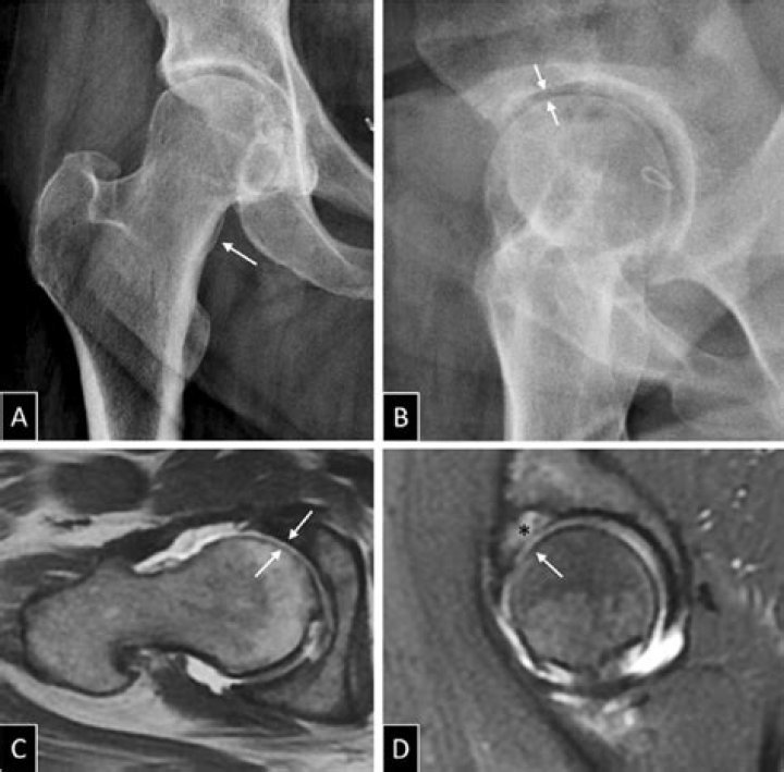
What is a Sublabral sulcus hip?
A posteroinferior sublabral groove is a relatively common normal anatomic hip variation. If not recognized as normal, the sulcus may serve as a diagnostic pitfall on MR arthrography. Its location is distinct from most labral tears.
What is the difference between arthrography and arthroscopy?
Whereas the arthrogram reproduces a black-and-white, two-dimensional picture of a spatial structure (an indirect procedure), arthroscopy provides a colored picture facilitating a three-dimensional assessment of the joint cavity, palpation, and the arthroscopic operation.
Does a hip MRI show the groin?
MRI is sensitive in diagnosing pathology in groin pain, with injuries to the adductor tendon attachment to the pubic tubercle most commonly identified. Not only can MRI be used to image rectus abdominis/adductor longus aponeurosis and pubic bone pathology, it can also identify hip or inguinal canal abnormalities.
Does your whole body go in for a hip MRI?
Often, an MRI will be targeted to a particular area of the body. If your hips are the area in question, a pelvic MRI will be performed. Pelvic MRIs allow a doctor to see the area between your hips, your reproductive organs, blood vessels, and hips themselves. Doctors will request hip MRIs for a variety of reasons.
What is Buford complex?
A Buford complex, found in 1.5% of individuals, is the absence of the anterior superior labrum in conjunction with a thickened cord-like middle glenohumeral ligament. A Buford complex can be confused with a sublabral foramen or pathologic labral detachment.
Can you drive after arthrogram and MRI of right hip?
After the procedure the joint will feel slightly unusual until the body has drained away all the injected fluid. We recommend that you do not drive or exercise for 24 hours after the procedure.
