What does increased T1 signal mean?
T1 weighted image – Pathology (spine) Loss of the normal high signal in the bone marrow indicates loss of normal fatty tissue and increased water content. Abnormal low signal on T1 images frequently indicates a pathological process such as trauma, infection, or cancer.
What is T1 in the brain?
T1 (longitudinal relaxation time) is the time constant which determines the rate at which excited protons return to equilibrium. It is a measure of the time taken for spinning protons to realign with the external magnetic field.
What is T1 hyperintense signal?
T1 signal hyperintensity may correspond to intracellular and extracellular methemoglobin. It may also be seen during the chronic stage of a clot or hemorrhage, when sedimentation of the blood cells produces a distinctive fluid-debris level within the lesion.
What is T1 hyperintense on MRI?
Hyperintense cerebral changes on T1-weighted images are formed due to accumulation of substances characterized by short longitudinal relaxation time including: gadolinium contrast, intra- and extracellular methemoglobin, melanin, fatty and protein-rich substances and minerals, i.a. calcium, copper and manganese.
What is T1 image in MRI?
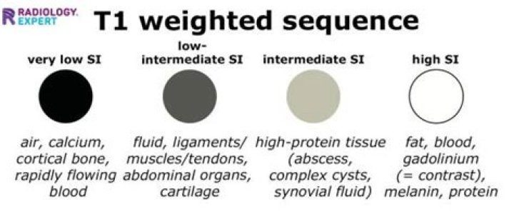
T1 weighted image (also referred to as T1WI or the “spin-lattice” relaxation time) is one of the basic pulse sequences in MRI and demonstrates differences in the T1 relaxation times of tissues. A T1WI relies upon the longitudinal relaxation of a tissue’s net magnetization vector (NMV).
What are T1 lesions?
T1 -hypointense lesions (T1-black holes) in multiple sclerosis (MS) are areas of relatively severe central nervous system (CNS) damage compared with the more non-specific T2-hyperintense lesions, which show greater signal intensity than normal brain on T2-weighted magnetic resonance imaging (MRI).
What is T1 weighted image?
What does T1 shortening mean?
Contrast enhanced The most commonly used contrast agents in MRI are gadolinium based. At the concentrations used, these agents have the effect of causing T1 signal to be increased (this is sometimes confusingly referred to as T1 shortening).
What causes T1 hyperintensity?
The main entities causing high T1 weighted signal in the basal ganglia include calcifications, haemorrhage and metal deposition. However, some disease processes such as infection and malignancy could give high T1 weighted signal due to one or a combination of factors such as haemorrhage and calcification.
What is a T1 and T2 hyperintense lesion?
T1 lesions were defined as regions with a signal intensity similar to or reduced to the signal intensity of gray matter and corresponding to a hyperintense region on T2-weighted MRI. Hyperintense–T2 lesions were defined as sharply demarcated regions of high signal intensity compared with surrounding brain tissue.
What does high signal on MRI mean?
High signal seen on these images indicates a pathological process such as infection, tumour, or areas of demyelination – as in this patient with multiple sclerosis.
