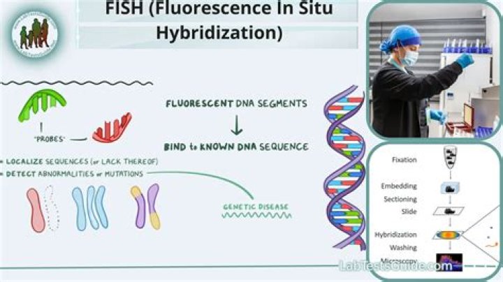
What does fluorescence in situ hybridization do?
Fluorescence in situ hybridization (FISH) provides researchers with a way to visualize and map the genetic material in an individual’s cells, including specific genes or portions of genes. This may be used for understanding a variety of chromosomal abnormalities and other genetic mutations.
How does RNA in situ hybridization work?
In situ hybridization is a laboratory technique in which a single-stranded DNA or RNA sequence called a probe is allowed to form complementary base pairs with DNA or RNA present in a tissue or chromosome sample. The probe has a chemical or radioactive label attached to it so that its binding can be observed.
What is mRNA hybridization?
Hybridization is a process whereby two single-stranded nucleic acid chains bind together by hydrogen bonding of complementary base pairs. ISH takes advantage of the ability of mRNA within a cell to hybridize with exogenously applied RNA or DNA molecules (Goodman and Spiegelman, 1971).
What does in situ hybridization detect?
In situ hybridization is a technique that is used to detect nucleotide sequences in cells, tissue sections, and even whole tissue. This method is based on the complementary binding of a nucleotide probe to a specific target sequence of DNA or RNA.
What does FISH test detect in pregnancy?
This testing allows preliminary detection of trisomy for chromosomes 13, 18, and 21, numerical abnormalities of the sex chromosomes, and triploidy (three sets of all chromosomes, resulting in 69 chromosomes). Prenatal interphase FISH can be performed on uncultured amniotic fluid, chorionic villi, or fetal blood cells.
What are the steps of in situ hybridization?
The major steps involved in in situ hybridization are as follows: probe preparation and labeling, tissue fixation, permeabilization, hybridization, and signal detection and these are described in detail in this chapter.
