What do abnormal P waves indicate?
What do abnormal P waves indicate?
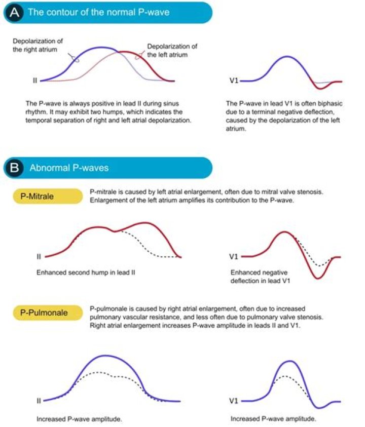
The Abnormal P wave If the p-wave is enlarged, the atria are enlarged. If the P wave is inverted, it is most likely an ectopic atrial rhythm not originating from the sinus node. Altered P wave morphology is seen in left or right atrial enlargement.
What does regularly irregular pulse mean?
An arrhythmia is an irregular heartbeat. It means your heart is out of its usual rhythm. It may feel like your heart skipped a beat, added a beat, or is “fluttering.” It might feel like it’s beating too fast (which doctors call tachycardia) or too slow (called bradycardia).
How do you calculate rate when rhythm is irregular?
SLOW or IRREGULAR rhythms:
- Rate = Number of R waves X 6.
- The number of complexes (count R waves) on the rhythm strip gives the average rate over a ten-second period. This is multiplied by 6 (10 seconds x 6 = 1 minute) to give the average Beats per minute (bpm)
What are P waves heart?
The P wave represents the electrical depolarization of the atria. In a healthy person, this originates at the sinoatrial node (SA node) and disperses into both left and right atria.
What is normal P axis in ECG?
The P wave is the first positive deflection on the ECG and represents atrial depolarisation. Normal P wave axis is between 0° and +75°.
Is irregular heartbeat always serious?
In many cases, these irregular heartbeats are harmless and will resolve on their own. But when they occur persistently, they can be serious. When your heart’s rhythm is disrupted, it isn’t pumping oxygenated blood efficiently, which can cause harm to the heart and the rest of the body.
Why is there no P wave in atrial fibrillation?
Because the atrial rate is so fast, and the action potentials produced are of such low amplitude, P waves will not be seen on the ECG in patients with atrial fibrillation.
How do you find P wave axis?
P-Wave Axis It is determined by measuring net positive or negative P-wave deflections on all six limb leads and calculating the net direction of electrical activity using the hexaxial reference system. Abnormal P-wave axis is defined as any value outside 0–75° (Figure 1) (31).
What is normal P wave in ECG?
P-wave checklist The P-wave is frequently biphasic in V1 (occasionally in V2). The negative deflection is normally <1 mm. P-wave duration should be ≤0,12 seconds. P-wave amplitude should be <2,5 mm in the limb leads.
What happens in P wave of ECG?
The P wave represents the depolarization of the left and right atrium and also corresponds to atrial contraction. Strictly speaking, the atria contract a split second after the P wave begins. Because it is so small, atrial repolarization is usually not visible on ECG.