What causes white spots on the cornea?
A corneal ulcer is most commonly caused by bacteria and fungal infections, though it can occur in anyone who has sustained a direct eye injury. The trauma creates an entry point for bacteria or other microorganisms to invade and establish an infection.
What does keratoconus look like on topography?
“Keratoconus is usually defined as a localized area of steepening on corneal topography,” notes Dr. Klyce. “This can occur anywhere on the corneal surface, and it has several faces. It can look like an asymmetric bow tie, for example.
How do you interpret corneal topography results?
Warmer colors represent steeper corneal curvature while cooler colors represent flatter areas. For the elevation maps (anterior and posterior float), warmer colors denote where the cornea is elevated above the best fit sphere and cooler colors denote where the cornea is depressed below the best fit sphere.
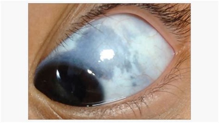
Why does my baby have a white spot on his eye?
A child that develops a white pupil or cloudy cornea needs immediate attention, preferably from an eye specialist. It is important to get diagnosed early if the problem is caused by retinoblastoma since this disease can be fatal.
Does keratoconus go away?
Keratoconus does not fade on its own. The shape of your cornea can’t permanently change, even with medications, special contact lenses, or surgery. Remember, we have various options for reshaping your cornea, but keratoconus is a chronic, lifelong disorder. So don’t wait until things get worse.
Is keratoconus serious?
Untreated keratoconus can lead to permanent vision loss. The changes to the cornea make it difficult for the eye to focus with or without eyeglasses or standard soft contact lenses.
How do you rule out keratoconus?
In order to make a diagnosis of keratoconus, the doctor must measure the curvature of the cornea. Several different tests can be performed to make the diagnosis. The test that is used most often is called topography. Topography measures the curvature of the surface of the eye and creates a colored “map” of the cornea.
How is keratoconus diagnosed?
Is keratoconus a value?
A value of 47.20 D or greater is suggestive of keratoconus. I-S value (inferior- superior value) quantifies the inferior versus superior corneal diopteric asymmetry that occurs in keratoconus. A positive value indicates higher inferior curvature while a negative value indicates higher superior curvature.
