What can be seen with electron microscope?
What can be seen with electron microscope?
An electron microscope is a microscope that uses a beam of accelerated electrons as a source of illumination. Electron microscopes are used to investigate the ultrastructure of a wide range of biological and inorganic specimens including microorganisms, cells, large molecules, biopsy samples, metals, and crystals.
What must be visually studied using electron microscopy?
The transmission electron microscope is used to view thin specimens (tissue sections, molecules, etc) through which electrons can pass generating a projection image. It provides detailed images of the surfaces of cells and whole organisms that are not possible by TEM.
Why can’t you view living things with an electron microscope?
Electron microscopes are the most powerful type of microscope, capable of distinguishing even individual atoms. However, these microscopes cannot be used to image living cells because the electrons destroy the samples.
What can an electron microscope see that a light microscope Cannot?
Electrons have much a shorter wavelength than visible light, and this allows electron microscopes to produce higher-resolution images than standard light microscopes. Electron microscopes can be used to examine not just whole cells, but also the subcellular structures and compartments within them.
What are 2 different types of electron microscopes?
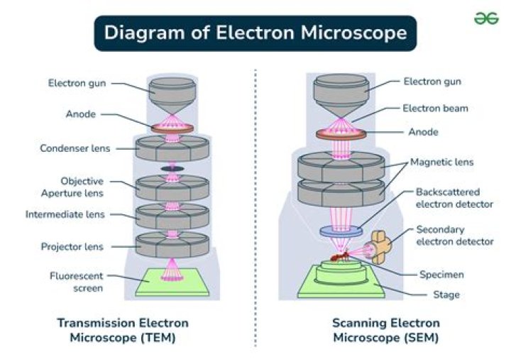
The two main types of electron microscopes are the transmission electron microscope (TEM) and the scanning electron microscope (SEM).
Can viruses be viewed on a light microscope?
Standard light microscopes allow us to see our cells clearly. However, these microscopes are limited by light itself as they cannot show anything smaller than half the wavelength of visible light – and viruses are much smaller than this.
What are the two types of electron microscopes?
What are the 3 types of electron microscopes?
There are several different types of electron microscopes, including the transmission electron microscope (TEM), scanning electron microscope (SEM), and reflection electron microscope (REM.)
What elements Cannot be detected with SEM?
EDS detectors on SEM’s cannot detect very light elements (H, He, and Li), and many instruments cannot detect elements with atomic numbers less than 11 (Na).
What is one advantage of a light microscope over an electron microscope?
light microscopy provides for higher resolving power than electron microscopy.
What has the highest magnification?
The highest magnification image ever created shows a single molecule of pentacene. Pentacene is a hydrocarbon which consists of five linearly fused benzene rings and has a molar mass of 278 g.
Why are electron microscopes bad for biological samples?
The electron beam inside a transmission electron microscope (TEM) causes problems for biological samples because of its high energy. It needs to have enough energy to pass right through the sample and out the other side.
What are examples of artefacts in electron microscope?
Micelles and strange-shaped mitochondria are examples of artefacts – structures that are seen under the microscope but aren’t found in living cells. It’s very important to be aware that artefacts can be introduced during fixation so that you don’t mistake them for real parts of your sample.
How are microtomes used in a scanning electron microscope?
Samples may be sectioned (with a microtome) if information about the organism’s internal ultrastructure is to be exposed for imaging. If the SEM is equipped with a cold stage for cryo microscopy, cryofixation may be used and low-temperature scanning electron microscopy performed on the cryogenically fixed specimens.
Why are living things cannot survive in an electron microscope?
Surviving a hostile environment. There are two reasons why living things can’t survive in an electron microscope: The power of the electron beam that’s directed at the sample. The vacuum inside the microscope.
What are the drawbacks of scanning electron microscopy?
Scanning electron microscopy (SEM) permits viewing of the surface of the specimen, and can produce a three-dimensional image. One major drawback to electron microscopy is that the process of preparing the specimen, as well as the actual process of examining the specimen, results in death of the cell or organism.
Can a sample be viewed on an electron microscope while alive?
However, these biological samples can’t be viewed on electron microscopes whilst alive. Instead, the samples must undergo complex preparation steps to help them withstand the environment inside the microscope. The preparation process kills the tissue and can also cause changes in the sample’s appearance.
Micelles and strange-shaped mitochondria are examples of artefacts – structures that are seen under the microscope but aren’t found in living cells. It’s very important to be aware that artefacts can be introduced during fixation so that you don’t mistake them for real parts of your sample.
What are the different types of electron microscopes?
There are two major types of electron microscopes used by scientists. Transmission electron microscopy (TEM) allows for viewing of the internal structures of the specimen being studied. Scanning electron microscopy (SEM) permits viewing of the surface of the specimen, and can produce a three-dimensional image.