What are the cells seen in blood film?
What are the cells seen in blood film?
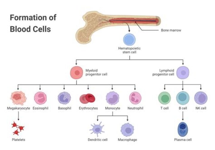
Red blood cells (RBCs) are the cells that transport oxygen to the tissues. White blood cells (WBCs) are cells that fight infection among several other functions. Platelets are cell fragments that play an important role in blood clotting.
What does film mean in a blood test?
A blood film is a snapshot of the cells that are present in the blood at the time that the sample is obtained. To produce a blood film, a single drop of blood is spread in a thin layer across a glass slide, dried, and then stained with a special dye.
What are the 3 kinds of cells you can see in the blood smear?
Blood Smear
- Red blood cells, which carry oxygen from your lungs to the rest of your body.
- White blood cells, which fight infection.
- Platelets, which help your blood to clot.
What is a normal blood film?
A blood smear is considered normal when your blood contains a sufficient number of cells and the cells have a normal appearance. A blood smear is considered abnormal when there’s an abnormality in the size, shape, color, or number of cells in your blood.
What do smudge cells indicate?
Smudge cells are remnants of cells that lack any identifiable cytoplasmic membrane or nuclear structure. Smudge cells, also called basket cells, are most often associated with abnormally fragile lymphocytes in disorders such as chronic lymphocytic leukemia (CLL).
What does 1+ Burr cells mean?
1+ means one quarter of cells are affected. 2+ means one half of cells are affected. 3+ means three quarters of cells are affected. 4+ means all of the cells are affected.
What is a film test?
noun. a filmed audition of a prospective actor or actress to test suitability.
How many types of blood films are there?
Two push-type peripheral blood smears suitable for characterization of cellular blood elements. Left smear is unstained, right smear is stained with Wright-Giemsa stain.
What teardrop cell means?
The presence of teardrop-shaped cells may indicate: Myelofibrosis. Severe iron deficiency. Thalassemia major. Anemia caused by bone marrow not producing normal blood cells due to toxins or tumor cells (myelophthisic process)
When do you see teardrop cells?
Teardrop cells (dacrocytes) are frequently associated with infiltration of the bone marrow by fibrosis, granulomatous inflammation, or hematopoietic or metastatic neoplasms. They can also be seen in patients with splenic abnormalities, vitamin B12 deficiency, and some other forms of anemia.
What is the importance of preparing a good blood film?
Inadequately prepared smear can present different artifacts and lead to errors in the differential count. Blood films should be made immediately after collection of the blood, because cell morphology deteriorates rapidly after sample collection.
What is the difference between a thick and thin blood film?
A thick blood smear is a drop of blood on a glass slide. A thin blood smear is a drop of blood that is spread across a large area of the slide.