How do you identify a myocardial infarction on an ECG?
The ECG findings of an acute anterior myocardial infarction wall include:
- ST segment elevation in the anterior leads (V3 and V4) at the J point and sometimes in the septal or lateral leads, depending on the extent of the MI.
- Reciprocal ST segment depression in the inferior leads (II, III and aVF).
Which of the ECG findings would be positive for an inferior wall MI?
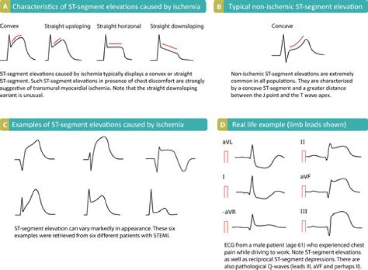
Upon ECG analysis, inferior STEMI displays ST-elevation in leads II, III, and aVF. There are subtle differences in the ECG pattern depending on the artery occluded. Reciprocal changes (ST-segment depression) may be seen in lead aVL [6].
Which leads are inferior MI?
Marked ST elevation in II, III and aVF with a “tombstone” morphology. Reciprocal change in aVL. ST elevation is also present in the lateral leads V5-6, indicating an extensive infarct of the inferior and lateral walls.
What are the inferior leads on ECG?
The arrangement of the leads produces the following anatomical relationships: leads II, III, and aVF view the inferior surface of the heart; leads V1 to V4 view the anterior surface; leads I, aVL, V5, and V6 view the lateral surface; and leads V1 and aVR look through the right atrium directly into the cavity of the …
What is an inferior infarction on ECG?
Inferior Wall ST Segment Elevation Myocardial Infarction (MI) ECG Review. An inferior wall MI — also known as IWMI, or inferior MI, or inferior ST segment elevation MI, or inferior STEMI — occurs when inferior myocardial tissue supplied by the right coronary artery, or RCA, is injured due to thrombosis of that vessel.
What does inferior infarct mean on ECG?
An inferior myocardial infarction (MI) is a heart attack or cessation of blood flow to the heart muscle that involves the inferior side of the heart. Inferior MI results from the total occlusion of either the right coronary artery in 85% of the cases or the left circumflex in 15% of the cases.
Where is an inferior MI?
Inferior wall myocardial infarction (MI) occurs from a coronary artery occlusion with resultant decreased perfusion to that region of the myocardium. Unless there is timely treatment, this results in myocardial ischemia followed by infarction.
How does inferior MI cause heart block?
Also, because the right coronary artery perfuses the sinoatrial node, heart block and bradycardia may occur. A high degree heart block, defined as a second or third-degree block, is seen in 19% of patients with acute inferior wall MI. The amount of collateral circulation to the AV impacts the rate of heart blocks.
What is meant by an inferior MI?
An inferior wall MI — also known as IWMI, or inferior MI, or inferior ST segment elevation MI, or inferior STEMI — occurs when inferior myocardial tissue supplied by the right coronary artery, or RCA, is injured due to thrombosis of that vessel.
How do you treat inferior myocardial infarction?
While heart blocks are a main contributor to morbidity and mortality, most high-degree heart blocks are treatable with atropine. It is seldom necessary to use a temporary pacemaker. The damaged myocardium can lead to potentially lethal arrhythmias such as ventricular tachycardia and ventricular fibrillation.
What is inferior myocardial?
