Does syphilis cause caseous necrosis?
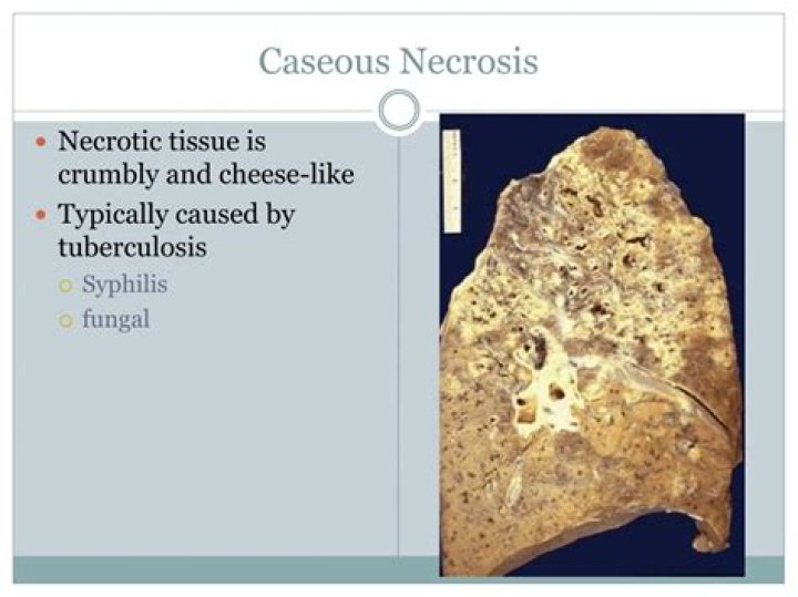
Frequently, caseous necrosis is encountered in the foci of tuberculosis infections. It can also be caused by syphilis and certain fungi. A similar appearance can be associated with histoplasmosis, cryptococcosis, and coccidioidomycosis.
What do Gummas look like?
A gumma is caused by the bacteria that cause syphilis. It appears during late-stage tertiary syphilis. It most often contains a mass of dead and swollen fiber-like tissue. It is most often seen in the liver.
What is caseous necrosis?
Caseous necrosis is a condition of cellular death that usually occurs in the lungs. When the lung cells die, the lungs take on a crumbly, dull-white appearance that resembles cheese. Although caseous necrosis most often occurs in the lungs, it can also happen in other locations of the body such as the kidneys.
When does caseous necrosis occur?
The caseous necrosis is the basic process of tuberculosis disease in humans. The interval from infection to tuberculin conversion is never more than 8 weeks and in general is 5 to 7 weeks (21). The onset of caseous necrosis coincides with the development of acquired immune resistance or CMI and DTH.
Does syphilis have granuloma?
Gummatous syphilis is characterized by granulomatous lesions, called gummas, which are characterized by a center of necrotic tissue with a rubbery texture. Gummas principally form in the liver, bones, and testes but may affect any organ.
Does syphilis cause Caseating granuloma?
Granulomatous inflammation is a hallmark of tertiary syphilis, which also presents with massive caseating necrosis [10-13]. Though uncommon, a prominent histiocytic inflammatory infiltrate has been described in lesions of secondary syphilis as well [11,14-17].
How do syphilis sores look like?
The rash can show up when your primary sore is healing or several weeks after the sore has healed. The rash can look like rough, red, or reddish brown spots on the palms of your hands and/or the bottoms of your feet. The rash usually won’t itch and it is sometimes so faint that you won’t notice it.
What is caseous lesion?
Caseous lesions consist of necrotic cellular debris surrounded by a zone of suppurative inflammation. Depending on the duration of the lesions, they may be partially encapsulated by fibrous tissue.
Where is Liquefactive necrosis most often seen?
Liquefactive necrosis usually occurs in the brain and results in a pus-filled cyst forming. Liquefactive necrosis most often occurs in the brain because the brain has a very high concentration of lysosomes.
What is Caseating granuloma?
Caseating granuloma means necrosis involving dead cells with no nuclei and debris. Without microscope, the cheese like pattern was seen in the these granulomas . In all reports of the CREMO patients, the granulomas were noncaseating .
What is syphilis reactive?
Interpretation of reactive tests A reactive treponemal test most likely indicates infection by T pallidum but is not sufficient to determine disease activity and make treatment decisions (table 1). A reactive test can be seen in patients with a history of syphilis who has been treated.
