Can MRI diagnose slap tear?
An MRI scan is often done to diagnose a SLAP tear and other potential injuries to the muscles, tendons, ligaments, and cartilage in the shoulder. Because of the many overlapping and interwoven structures in the shoulder, it is possible for an MRI scan to miss a smaller tear.
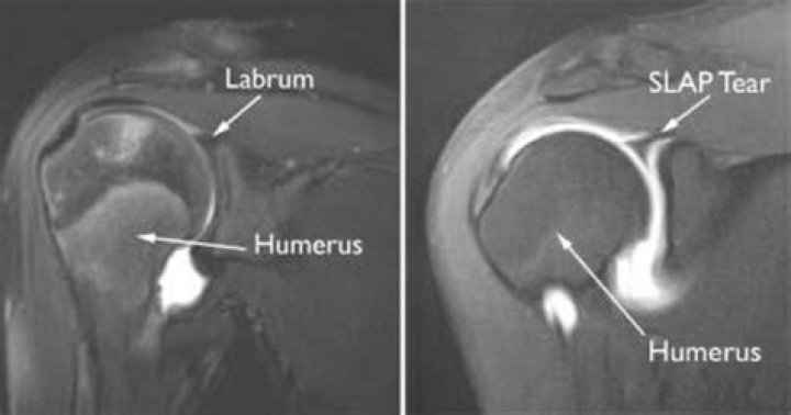
How do you check for SLAP tear in MRI?
Common diagnostic criteria for a SLAP lesion by MR or MR arthrography include the following: presence of a laterally curved, high signal intensity in the labrum on a coronal image, multiple or branching lines of high signal intensity in the superior labrum on a coronal image, full-thickness detachment with irregularly …
Will an MRI show a shoulder labral tear?
Diagnosing a labrum tear involves a physical examination and most likely an MRI, CT scan and/or arthroscopy of the shoulder. Treatment varies depending on type, severity and location of the labrum tear.
What is the difference between an MRI and MRI arthrogram?
MRI provides a detailed look at most body structures including soft tissues. An Arthrogram uses fluoroscopy and an MRI to specifically diagnoses injuries in the joint structures that an MRI alone would likely miss.
Is a SLAP tear the same as a labrum tear?
A SLAP tear is also referred to as a labral tear, or a tear or lesion to the labrum. This injury tends to develop over time due to repetitive movements. It can also result from acute trauma or age.
What is MRI arthrogram shoulder?
What is an MR Arthrogram? An arthrogram uses imaging equipment to evaluate a joint like the shoulder, elbow, wrist, hip, knee or ankle. It is a two-part procedure consisting of a contrast injection into the joint, followed by an MRI or CT scan of the joint.
What does an arthrogram of the shoulder show?
An arthrogram is a test that helps healthcare providers diagnose joint problems, such as hip or shoulder pain. MR arthrograms can show ligament, tendon and cartilage issues with clear detail. You get a special dye injected into your joint before having an MRI scan or other imaging test.
What does an MRI arthrogram of the shoulder show?
It is a two-part procedure consisting of a contrast injection into the joint, followed by an MRI or CT scan of the joint. An arthrogram is ordered to: Find tears, degeneration or disease in the cartilage, ligament or tendon. Detect growths or synovial cysts in the joint.
What does an MRI arthrogram show?
What can a shoulder arthrogram show?
For the shoulder, an arthrogram may be requested by your doctor for the following reasons: Suspected tear of the labrum or cartilage lining of the joint. To show whether a tear of the tendons of the rotator cuff is full-thickness, meaning going all the way through the tendon. To evaluate the shoulder after surgery.
Is arthrogram of shoulder painful?
While the arthrography procedure itself causes no pain, having to move or hold the joint still in certain positions might cause some discomfort or pain, particularly if you’ve recently had surgery or a joint injury.
What is an arthrogram MRI of the shoulder?
